The parts of the foot work together every time you stand, walk, run, jump, climb stairs, or simply shift your weight from one side to another. Although the foot may look like a fairly simple structure, it is actually a highly organized combination of bones, joints, muscles, tendons, ligaments, nerves, blood vessels, and soft tissues. Each component has a specific role, and even a small problem in one area can affect how the entire foot functions.
Understanding foot anatomy can make it much easier to recognize why certain areas hurt or become uncomfortable. For example, pain beneath the heel may have a different cause from pain in the ball of the foot, while discomfort around the ankle can involve structures that connect the lower leg to the foot. Knowing the basic parts of the foot also helps you understand common conditions, footwear issues, sports injuries, and everyday foot care.
In this guide, we will explore the major parts of the foot from the heel and arch to the toes, while also looking at the bones, joints, muscles, tendons, ligaments, nerves, and other structures hidden beneath the skin. The goal is not to make anatomy complicated. Instead, we will break it down into practical sections so you can understand what each part does and why it matters.
What Are the Main Parts of the Foot?
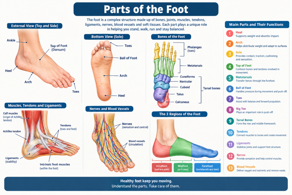
The human foot is commonly divided into three broad regions: the hindfoot, midfoot, and forefoot. These areas are connected rather than completely separate, and they work together to provide support, balance, flexibility, and movement. The hindfoot includes the heel region and important ankle-related bones, the midfoot contains the arch-forming structures, and the forefoot includes the ball of the foot and toes.
Another simple way to understand the foot is by looking at its visible areas. The heel is at the back, the sole is underneath, the top of the foot is called the dorsum, and the toes extend from the front. The medial side is the side facing toward the body’s center, while the lateral side is the outer edge. These terms are commonly used by doctors and other healthcare professionals when describing foot problems.
Underneath these recognizable areas is a complex framework of bones and connective tissues. There are 26 bones in one human foot, along with numerous joints and many muscles and tendons. This combination allows the foot to be both strong enough to carry body weight and flexible enough to adapt to different surfaces.
The Heel: One of the Most Important Parts of the Foot
The heel is the rear portion of the foot and is one of the first areas to receive significant force when many people walk. The main bone forming the heel is the calcaneus, which is the largest bone in the foot. It provides a strong foundation for weight bearing and connects with other bones that help form the rear part of the foot.
A thick layer of cushioning tissue beneath the heel helps absorb impact. Every step can place considerable force through the foot, particularly during running or jumping, so this natural padding plays an important protective role. The heel also provides an attachment point for the Achilles tendon, one of the body’s strongest tendons.
Heel discomfort can develop for many reasons, including overuse, unsuitable footwear, injuries, or irritation of nearby tissues. Plantar fasciitis is one well-known cause of heel pain, although several other conditions can also produce similar symptoms. Persistent or severe heel pain should not automatically be blamed on one specific condition without an appropriate evaluation.
The Arch of the Foot
The arch is one of the most recognizable structural features among the parts of the foot. It runs along the inside of the foot and helps distribute body weight while also contributing to shock absorption. The arch is not simply an empty space beneath the foot; it is supported by bones, ligaments, muscles, tendons, and connective tissue.
Most people think primarily about the medial longitudinal arch, which runs along the inner side of the foot. However, the foot also has a lateral longitudinal arch and a transverse arch. Together, these structures create a flexible platform that can adjust as the foot moves across different surfaces.
Arch shape varies naturally from person to person. Some people have relatively low arches, while others have high arches. Neither shape automatically means that a person has a medical problem. What matters more is how the foot functions, whether symptoms are present, and whether the structure contributes to abnormal stress or movement.
The Sole of the Foot
The sole is the underside of the foot, the part that comes into direct contact with the ground during standing and walking. It is also known anatomically as the plantar surface. Because this surface experiences repeated pressure, it has specialized skin and cushioning tissues designed to tolerate everyday mechanical stress.
The skin on the sole is generally thicker than the skin on many other parts of the body. Beneath it is a layer of fatty tissue that helps provide cushioning. This padding is particularly useful beneath areas that experience substantial pressure, such as the heel and the ball of the foot.
The sole also contains important structures involved in movement and sensation. Muscles, tendons, nerves, and connective tissues work together beneath the foot. Problems affecting these structures can cause pain that becomes noticeable while walking, standing for long periods, or exercising.
The Top of the Foot
The top of the foot is called the dorsum or dorsal surface. Unlike the sole, the skin on the top is generally thinner and has less natural cushioning. Several tendons responsible for lifting and extending the toes pass across this area.
The bones underneath the top of the foot include structures from the tarsal and metatarsal regions. These bones contribute to the framework that supports the foot and transfers forces between the ankle and the toes. Because the bones and tendons are relatively close to the surface, swelling or irritation in this region can sometimes be easy to notice.
Pain on top of the foot may have different causes. Tight footwear can place pressure on the tendons and bones, while repetitive activity can contribute to overuse injuries. Trauma, stress injuries, inflammation, or problems involving the joints may also produce discomfort in this area.
The Bones in the Foot
One of the most important facts about the parts of the foot is that each foot contains 26 bones. These bones are grouped into three major categories: the tarsal bones, metatarsals, and phalanges. Their arrangement creates a framework that is strong but still capable of controlled movement.
The seven tarsal bones form much of the rear and middle portion of the foot. They include the talus and calcaneus, along with the navicular, cuboid, and three cuneiform bones. Farther forward are the five metatarsals, which connect the midfoot with the toes.
The toes contain the phalanges. Four of the lesser toes have three phalanges each, while the big toe normally has two. Although these bones are relatively small, they are important during walking because the toes help stabilize the body and assist with the push-off phase of each step.
The Tarsal Bones
The tarsal bones form the back and middle sections of the foot. The calcaneus is the heel bone, while the talus sits above it and forms an important connection with the bones of the lower leg. The talus plays a particularly important role in transferring body weight from the ankle into the foot.
The remaining tarsal bones include the navicular, cuboid, and three cuneiforms. Their arrangement contributes to the shape and stability of the midfoot. These bones are connected by joints and strong ligaments, allowing controlled movement while preventing excessive instability.
Because the tarsal region carries substantial mechanical loads, injuries here can interfere with normal walking. Some injuries are obvious after a major accident, while others can develop gradually from repetitive stress. Proper assessment is important when pain persists or is associated with swelling, bruising, or difficulty bearing weight.
The Metatarsals
The five metatarsal bones occupy the middle-to-front portion of the foot. They are numbered from the medial side outward, with the first metatarsal associated with the big toe and the fifth associated with the little toe. These long bones form an important bridge between the tarsal bones and the toe bones.
The metatarsals help distribute pressure across the forefoot. During walking, forces move through these bones as the body progresses forward. The first metatarsal and the structures surrounding the big toe are especially important during the final push-off stage.
Metatarsal problems can occur after direct trauma or repetitive loading. Stress injuries are particularly relevant for people who suddenly increase their activity level. Footwear, training surfaces, biomechanics, and individual anatomy can all influence how pressure is distributed across the metatarsal region.
The Toes and Phalanges
The toes are the frontmost parts of the foot and are important for balance and forward movement. The big toe is known as the hallux, while the remaining toes are often described as the second through fifth toes. Each toe has a specific position and contributes to the overall function of the forefoot.
The four smaller toes usually contain three phalanges: proximal, middle, and distal. The big toe contains only proximal and distal phalanges. These small bones are connected through joints and supported by muscles, tendons, and ligaments.
Toes can experience a variety of problems, including blisters, corns, calluses, nail disorders, fractures, and deformities. Shoes that are too narrow or short can place repeated pressure on the toes. Choosing footwear with adequate length and room around the forefoot can reduce unnecessary mechanical pressure.
The Big Toe and Why It Matters
The big toe may be small compared with the rest of the foot, but it plays a major role in movement. During normal walking, the big toe helps provide a stable point for push-off. It also contributes to balance and helps the foot transition from a flexible structure to a more rigid lever during the later stage of a step.
The joint at the base of the big toe is called the first metatarsophalangeal joint. This joint needs sufficient movement for efficient walking. Problems in this area can change how a person distributes pressure across the rest of the foot.
One familiar condition affecting the big toe is a bunion, which involves a structural change around the base of the big toe and can produce a visible bump. Other problems include arthritis, injuries, and stiffness. Because the big toe has such an important mechanical role, persistent symptoms can influence the way the entire lower limb moves.
The Ball of the Foot
The ball of the foot refers to the padded area beneath the forefoot, just behind the toes. Anatomically, this region is associated with the heads of the metatarsal bones and surrounding soft tissues. It carries considerable pressure during walking, running, and other activities.
The ball of the foot helps transfer body weight toward the toes. During the push-off portion of a stride, pressure can increase significantly in this area. The natural fat pads beneath the forefoot provide cushioning and help protect deeper structures from repetitive impact.
Pain in the ball of the foot is sometimes described as metatarsalgia, although that term describes a symptom pattern rather than a single specific cause. Activity, footwear, foot structure, injuries, and altered pressure distribution can all contribute to discomfort in this region.
Muscles of the Foot
Muscles are essential parts of the foot because they help control movement and stability. Some muscles are located within the foot itself, while others originate in the lower leg and send tendons into the foot. Together, they control the toes and help maintain the shape and function of the arches.
Intrinsic foot muscles are located within the foot. They assist with movements of the toes and help stabilize the foot during weight bearing. Although these muscles are not as large as the muscles of the thigh or calf, their coordinated activity is important for efficient movement.
The muscles of the lower leg also contribute significantly to foot movement. For example, muscles in the front of the lower leg help lift the foot and toes, while muscles in the back help push the foot downward. Their tendons connect these muscles to bones in the foot.
Tendons in the Foot
Tendons are strong bands of connective tissue that attach muscles to bones. Several important tendons cross the ankle and enter the foot, allowing muscles in the lower leg to control foot and toe movements.
The Achilles tendon connects the calf muscles to the calcaneus. It plays a major role in pushing the foot downward during activities such as walking, running, and jumping. Other tendons around the ankle help control inversion, eversion, dorsiflexion, and toe movement.
Tendon irritation can develop when a structure is repeatedly loaded beyond its ability to recover. Sudden increases in exercise, repetitive movements, poor biomechanics, and inadequate recovery can contribute to tendon problems. Gradual training and appropriate footwear can help reduce unnecessary stress.
Ligaments and Foot Stability
Ligaments are tough bands of connective tissue that connect bones to other bones. They are among the key parts of the foot responsible for maintaining joint stability. Without ligaments, the bones would not have the same controlled relationship during movement.
There are numerous ligaments throughout the foot and ankle. Some support individual joints, while others help maintain the arches. The plantar ligaments underneath the foot are particularly important for supporting the structure during weight bearing.
Ligament injuries can range from mild stretching to more significant tears. An ankle sprain, for example, can affect ligaments around the ankle and may temporarily alter how forces are distributed through the foot. Recovery depends on the severity of the injury and the structures involved.
Joints of the Foot
Joints are the places where bones meet, and the foot contains many of them. These joints allow the foot to bend, rotate, adapt to surfaces, and create the controlled movements required for walking.
The ankle joint is particularly important because it connects the lower leg with the foot. Beneath and around it are joints involving the talus, calcaneus, navicular, cuboid, and other bones. The smaller joints between the metatarsals and phalanges allow the toes to move.
Healthy joints require appropriate alignment, cartilage, connective tissues, and surrounding muscles. Problems such as arthritis, injuries, inflammation, or structural changes can reduce mobility and cause pain. Joint symptoms that persist should be evaluated rather than ignored.
Nerves in the Foot
Nerves provide sensation and help control muscles. They allow the brain to receive information about pressure, temperature, touch, and pain from the foot. This sensory information is important because it helps the body respond to changes in the ground and maintain balance.
Several major nerves travel from the lower leg into the foot and divide into smaller branches. These branches supply different regions of the sole, top of the foot, and toes. Because nerves can be compressed or irritated, nerve-related foot symptoms may include burning, tingling, numbness, or unusual sensitivity.
Nerve symptoms deserve attention, particularly when they persist or progressively worsen. A person experiencing unexplained numbness, weakness, or significant changes in sensation should seek appropriate medical advice to identify the underlying cause.
Blood Vessels and Circulation in the Foot
The foot needs a reliable blood supply to deliver oxygen and nutrients to its tissues. Arteries carry blood toward the foot, while veins return blood toward the heart. Smaller blood vessels distribute blood throughout the skin, muscles, bones, and other tissues.
Good circulation supports normal tissue health and healing. When circulation is reduced, wounds may take longer to heal and tissues can become more vulnerable to injury. This is one reason foot health deserves particular attention in people who have conditions that can affect circulation.
Changes such as persistent swelling, unusual color, temperature differences, or wounds that do not heal should not be ignored. These symptoms can have many possible explanations, so professional assessment may be appropriate when they persist or appear suddenly.
Why the Foot Has Arches
The arches of the foot are designed to combine flexibility with support. They help the foot adapt to the ground while distributing mechanical forces across different structures. Rather than acting like rigid beams, the arches respond dynamically as the body moves.
When the foot accepts weight, the arches can flatten slightly and then return toward their resting position. This controlled movement helps with shock absorption and adaptation. During push-off, the foot becomes more rigid so it can act as an effective lever.
Arch function depends on many structures rather than a single bone or muscle. Bones provide the framework, ligaments provide passive support, and muscles and tendons provide active control. This combination is what makes the foot both stable and mobile.
How the Parts of the Foot Work Together When You Walk
Walking may feel effortless, but it requires coordinated movement throughout the foot and lower limb. As the foot contacts the ground, different areas progressively accept and transfer body weight. The heel, midfoot, forefoot, and toes each contribute to this process.
The foot needs to adapt when it first contacts the ground. As the body moves forward, the midfoot helps manage forces and accommodate the surface. Later, the forefoot and toes become increasingly important as the body prepares to move into the next step.
This sequence demonstrates why an issue in one area can sometimes affect another. If a painful toe changes the way someone walks, for example, additional stress may be placed on other areas. The body naturally compensates, but prolonged compensation can sometimes contribute to new discomfort.
Common Problems Affecting the Parts of the Foot
Foot problems can affect virtually any structure. Common complaints include heel pain, arch discomfort, pain in the ball of the foot, toe problems, blisters, calluses, and injuries. Some conditions are temporary, while others require ongoing management.
Plantar fasciitis is commonly associated with pain around the heel and bottom of the foot. Bunions affect the area around the base of the big toe, while conditions such as hammertoe can change the position of a toe. Fractures and sprains can affect bones and ligaments after injuries.
Not every episode of foot pain means something serious. However, pain that is severe, persistent, associated with significant swelling or deformity, or preventing normal weight bearing deserves attention. Getting the correct diagnosis is more useful than simply treating the location of pain without understanding its cause.
How Footwear Affects Foot Anatomy
Shoes influence how pressure is distributed across the parts of the foot. A well-fitting shoe should provide enough length and width for the foot to sit comfortably without excessive compression. The shape of the shoe should also accommodate the natural shape of the forefoot.
Shoes that are too narrow can squeeze the toes together and increase pressure around the forefoot. Shoes with inadequate cushioning may feel uncomfortable during prolonged activity, while excessively soft or unstable footwear may not provide the support some people need.
The best footwear depends on the individual and the activity. Walking shoes, running shoes, work footwear, and sports shoes have different design goals. Rather than choosing shoes solely because they look good, it is sensible to consider fit, comfort, intended activity, and the individual’s foot structure.
Basic Foot Care Tips
Taking care of your feet does not have to be complicated. Washing and drying the feet regularly, keeping the skin appropriately moisturized, and choosing comfortable footwear are simple habits that can support overall foot health. Toenails should also be trimmed carefully and regularly.
It is useful to check the feet occasionally for blisters, cuts, swelling, irritation, changes in skin appearance, or areas of unusual pressure. Small problems can become more troublesome if they are repeatedly irritated by shoes or activity.
People who have reduced sensation or circulation may need to be particularly careful with routine foot checks. When sensation is reduced, an injury may not be felt immediately. In such situations, professional guidance about foot care can be especially valuable.
When Should You See a Doctor About Foot Pain?
Occasional soreness after unusual activity may improve with rest and basic self-care. However, persistent or recurring pain is worth investigating, particularly if it interferes with walking, work, sleep, exercise, or daily activities.
Medical attention is especially important after a significant injury, when the foot appears deformed, when you cannot comfortably bear weight, or when swelling and bruising are substantial. Numbness, weakness, unexplained color changes, or a wound that does not heal are also reasons to seek professional evaluation.
A healthcare professional can examine the painful area and consider how the different parts of the foot are functioning together. Depending on the situation, evaluation may include movement testing, imaging, or other diagnostic approaches. Treatment should be based on the underlying problem rather than simply the location of discomfort.
Quick Guide to the Main Parts of the Foot
| Part of the foot | Main role |
|---|---|
| Heel | Supports weight and absorbs impact |
| Arch | Helps distribute weight and adapt to surfaces |
| Sole | Provides contact, traction, cushioning, and sensation |
| Top of foot | Contains bones and tendons involved in movement |
| Metatarsals | Transfer forces through the forefoot |
| Ball of foot | Handles pressure during movement and push-off |
| Toes | Assist with balance and forward propulsion |
| Big toe | Plays an important role in push-off |
| Tarsal bones | Form the rear and middle framework |
| Tendons | Connect muscles to bones and create movement |
| Ligaments | Stabilize joints and support foot structure |
| Nerves | Provide sensation and help control muscles |
Final Thoughts on the Parts of the Foot
The parts of the foot may be small compared with the rest of the body, but they perform an impressive range of jobs. Bones provide structure, joints allow movement, ligaments create stability, tendons transfer muscular force, nerves provide sensation, and soft tissues help cushion repeated impact. None of these structures works entirely alone. The heel helps manage impact, the arches distribute forces, the forefoot transfers pressure, and the toes contribute to balance and push-off. Together, these areas allow the foot to adapt to uneven surfaces while supporting the body during everyday activities.
Understanding foot anatomy can also make it easier to pay attention to changes before they become major problems. Comfortable footwear, sensible activity progression, routine foot care, and appropriate medical attention when symptoms persist can all contribute to healthier feet. When you understand how the different structures work together, it becomes much easier to appreciate just how sophisticated the human foot really is.
Frequently Asked Questions About the Parts of the Foot
How many bones are in one foot?
Each human foot normally contains 26 bones. These are divided into seven tarsal bones, five metatarsals, and fourteen phalanges. Together, these bones form the structural framework of the foot.
What are the three main sections of the foot?
The foot is commonly divided into the hindfoot, midfoot, and forefoot. The hindfoot includes the heel and talus region, the midfoot contains several bones that contribute to the arches, and the forefoot includes the metatarsals and toes.
What is the bottom of the foot called?
The bottom of the foot is called the plantar surface or sole. It contains specialized skin, cushioning tissue, muscles, tendons, nerves, and other structures that help the foot handle pressure and movement.
What is the top of the foot called?
The top of the foot is called the dorsum or dorsal surface. Several tendons, bones, nerves, and blood vessels are located in or around this region.
Why is the big toe important?
The big toe is particularly important for balance and propulsion. During walking, it helps the foot create a stable lever during the push-off phase, making it an important part of efficient movement.
What supports the arch of the foot?
The arch is supported by a combination of bones, ligaments, muscles, tendons, and connective tissues. No single structure is solely responsible for maintaining the arch.
Why does the ball of my foot hurt?
Pain in the ball of the foot can have several causes, including excessive pressure, footwear issues, repetitive activity, injuries, and changes in foot mechanics. Persistent pain should be evaluated to determine the underlying cause.
Can shoes change how the foot functions?
Yes. Footwear can influence pressure distribution, toe position, cushioning, stability, and overall comfort. Shoes that do not fit properly may increase pressure on particular areas of the foot.
Are flat feet always a problem?
No. A low arch does not automatically mean that something is wrong. Many people have naturally flatter feet without experiencing pain or functional limitations. Symptoms and foot function are more important than arch height alone.
What is the strongest tendon associated with the foot?
The Achilles tendon is one of the strongest tendons in the body. It connects the calf muscles to the heel bone and plays an important role in walking, running, jumping, and pushing the foot downward.
How can I keep my feet healthy?
Regular hygiene, appropriate footwear, gradual increases in physical activity, routine inspection of the feet, and attention to persistent symptoms are useful foundations for foot health. If you develop ongoing pain or significant changes, professional evaluation can help identify the cause.
YOU MAY HAVE MASSED